

Coronary Angiography
Coronary angiography in Turkey is an invasive diagnostic procedure used to visualize the coronary arteries and identify narrowing or blockages that may reduce blood flow to the heart. It is commonly performed when coronary artery disease is suspected, when symptoms require further investigation, or when previous cardiac tests suggest significant arterial narrowing. The procedure can also help determine whether medical treatment, angioplasty, stent placement, or coronary bypass surgery may be appropriate.

What Is Coronary Angiography?
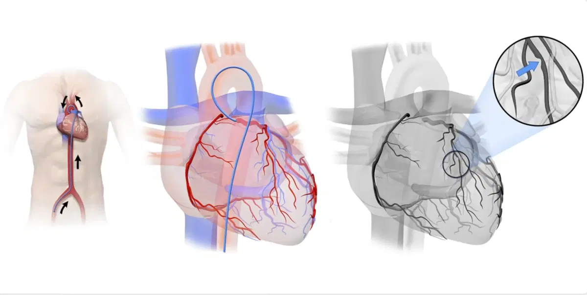
Coronary angiography is an imaging procedure that shows the arteries supplying blood to the heart muscle. A thin catheter is introduced into an artery and guided toward the heart. Contrast material is then injected into the coronary circulation. X-ray imaging records how the contrast moves through the arteries. This allows the cardiology team to identify areas where blood vessels are narrowed or completely blocked. Coronary angiography is considered different from routine non-invasive cardiac imaging because it directly examines the coronary artery lumen through catheter-based contrast imaging.
Why Is Coronary Angiography Performed?
Coronary angiography is usually performed when more detailed information about coronary blood flow is required.
It may be recommended for patients with persistent chest pain, suspected angina, abnormal stress test results, or signs of reduced blood supply to the heart.
The procedure is also commonly used during the evaluation of acute coronary syndromes and heart attacks.
Patients preparing for selected heart valve or cardiovascular operations may undergo angiography to determine whether significant coronary artery disease is also present.
What Is Coronary Artery Disease?
Coronary artery disease develops when plaque accumulates inside the arteries that supply the heart.
This process, known as atherosclerosis, can gradually narrow the arterial lumen and reduce blood flow.
Patients may experience chest pressure, shortness of breath, reduced exercise tolerance, or other symptoms.
A plaque can also rupture and trigger sudden blood clot formation.
If the clot significantly obstructs coronary blood flow, it can cause a heart attack.
Coronary angiography can identify the location and severity of these obstructions.
How Is Coronary Angiography Performed?
The procedure is usually performed in a cardiac catheterization laboratory.
The access area is cleaned and local anesthesia is used before a catheter is inserted into an artery.
The catheter is then guided through the vascular system toward the coronary arteries.
Contrast dye is injected while X-ray images are recorded from different angles.
The cardiologist evaluates the coronary anatomy and measures the significance of visible narrowing.
If an appropriate blockage is identified, angioplasty or stenting may sometimes be performed during the same session.
What Is Radial Coronary Angiography?
Radial angiography uses an artery in the wrist as the catheter entry point.
This approach is widely used because it can allow earlier mobilization after the procedure.
Bleeding complications at the access site may also differ compared with femoral access in selected patients.
Before the procedure, circulation to the hand and the suitability of the radial artery can be assessed.
Radial access may not be appropriate in every case, particularly when vascular anatomy, previous procedures, or technical requirements make another access route preferable.
What Is Femoral Coronary Angiography?
Femoral coronary angiography uses an artery in the groin as the access route.
The femoral artery provides a relatively large pathway for cardiac catheters and remains useful for selected diagnostic and interventional procedures.
After the catheter is removed, pressure or a vascular closure device may be used to control bleeding.
Patients generally need to limit movement of the treated leg for a period after the procedure.
The choice between radial and femoral access depends on clinical condition, vascular anatomy, procedure complexity, and operator assessment.
What Does Coronary Angiography Show?
Coronary angiography shows the location, length, and apparent severity of narrowing within the coronary arteries.
It also provides information about complete arterial blockages, branching anatomy, collateral circulation, and previous stents or bypass grafts.
The findings can help determine whether a lesion is technically suitable for catheter-based treatment.
However, angiography primarily shows the inside outline of blood vessels.
Additional techniques may sometimes be needed to understand whether a moderate narrowing significantly restricts blood flow or to assess plaque characteristics more precisely.
What Are FFR and iFR?
Fractional Flow Reserve, or FFR, and instantaneous wave-free ratio, or iFR, are techniques used to assess whether a coronary narrowing significantly affects blood flow.
They can be particularly useful when angiography shows an intermediate narrowing whose clinical importance is uncertain.
A pressure-sensitive wire is passed across the lesion to compare pressure before and after the narrowed area.
These measurements can help determine whether revascularization is likely to be beneficial.
They are not required for every coronary lesion and are used according to the clinical situation.
What Are IVUS and OCT?
Intravascular Ultrasound, or IVUS, and Optical Coherence Tomography, or OCT, provide images from inside the coronary artery.
IVUS uses ultrasound, while OCT uses light-based imaging.
These technologies can show vessel dimensions, plaque characteristics, and the relationship between a stent and the artery wall.
They may be used when angiography alone does not provide sufficient detail.
IVUS and OCT can also support the planning and optimization of complex coronary interventions.
Their use depends on lesion anatomy, procedural objectives, and clinical need.
Coronary Angiography vs CT Coronary Angiography
Invasive coronary angiography and CT coronary angiography both visualize coronary arteries but use different techniques.
CT coronary angiography is non-invasive and uses computed tomography with intravenous contrast.
It can be useful for evaluating selected patients with suspected coronary artery disease.
Conventional angiography requires arterial catheterization but offers the important advantage of allowing immediate treatment when appropriate.
The preferred test depends on symptoms, cardiovascular risk, previous test results, kidney function, heart rhythm, and the likelihood that an intervention will be required.
Can a Stent Be Placed During Coronary Angiography?
Yes, angioplasty and stent placement can sometimes be performed immediately after diagnostic coronary angiography.
If a significant treatable narrowing is identified, the cardiologist may proceed with Percutaneous Coronary Intervention, or PCI.
A balloon catheter can be used to expand the narrowed area before a stent is placed.
The stent helps maintain blood flow through the treated artery.
Whether immediate intervention is appropriate depends on coronary anatomy, symptoms, consent, medication status, and the overall treatment strategy.
When Is CABG Recommended After Angiography?
Coronary angiography may show disease that is more appropriately treated with Coronary Artery Bypass Grafting rather than stenting.
CABG can be considered for selected patients with extensive multivessel coronary disease, complex lesions, or significant disease involving important coronary segments.
Diabetes, heart function, age, and overall coronary anatomy can also influence the decision.
In complex cases, angiography findings may be reviewed by both interventional cardiologists and cardiovascular surgeons before choosing between medical treatment, PCI, and bypass surgery.
Is Coronary Angiography Painful?
Coronary angiography is generally performed using local anesthesia at the arterial access site.
Patients may feel brief pressure during catheter insertion or a temporary warm sensation when contrast material is injected.
The catheter moving inside the blood vessels is usually not felt in the same way as an external instrument.
Discomfort can occur at the wrist or groin access site after the procedure.
Sedative medication may be used in selected patients, but general anesthesia is not routinely required for standard diagnostic coronary angiography.
What Are the Risks of Coronary Angiography?
Coronary angiography is routinely performed but remains an invasive cardiovascular procedure.
Possible complications include bleeding, bruising, arterial injury, abnormal heart rhythm, allergic reactions to contrast material, and temporary kidney function changes.
More serious complications such as stroke, heart attack, severe vascular injury, or emergency surgery are less common.
Risk varies according to age, kidney function, cardiovascular condition, vascular disease, medications, and whether the procedure is diagnostic or combined with an intervention.
How Long Is Recovery After Coronary Angiography?
Recovery is usually relatively short after uncomplicated diagnostic angiography.
Patients are monitored for bleeding, blood pressure changes, heart rhythm problems, and access-site complications.
Those treated through the radial artery can often begin walking relatively early.
Femoral access may require a longer period of lying down to protect the puncture site.
Strenuous activity and heavy lifting may be temporarily restricted.
Recovery takes longer if angioplasty, stent placement, or another cardiovascular intervention is performed during the same session.
What Happens After Coronary Angiography?
The cardiologist reviews the angiography findings and develops a treatment strategy based on the coronary anatomy.
Patients without significant obstructive disease may continue with medical management and cardiovascular risk reduction.
Others may require cholesterol treatment, antiplatelet medication, blood pressure control, or diabetes management.
Significant coronary disease may lead to PCI or referral for bypass surgery.
Smoking cessation, physical activity, nutrition, and long-term cardiovascular follow-up remain important because angiography identifies disease but does not itself stop atherosclerosis.
Coronary Angiography in Turkey for International Patients
International patients considering coronary angiography in Turkey should provide previous cardiac records whenever available.
Useful documents may include ECG results, echocardiography reports, stress tests, CT coronary angiography, previous angiography images, stent records, blood tests, and current medication lists.
Patients using anticoagulants, antiplatelet drugs, or diabetes medication should ensure these treatments are reviewed before the procedure.
Travel planning should also consider the possibility that angiography may identify a condition requiring angioplasty, stenting, or cardiovascular surgery.
Coronary angiography in Turkey can provide detailed assessment of coronary artery anatomy and help guide treatment decisions for suspected or established coronary artery disease. Appropriate patient selection, careful arterial access, accurate interpretation of findings, and structured cardiovascular follow-up remain central to coronary care.

You can send us messages regarding your appointments and requests.
 Send Message